Introduction
The clinical success of GalNAc-conjugated siRNAs established a powerful paradigm for nucleic acid therapeutics (NATs): effective delivery through receptor-mediated endocytosis (RME) can transform a promising nucleic acid into a viable medicine. GalNAc succeeded because it leveraged the asialoglycoprotein receptor (ASGPR), which is highly expressed on hepatocytes and undergoes efficient receptor recycling. As a result, the liver has become the first organ in which systemic delivery of oligonucleotides can be achieved routinely and predictably.1 In contrast, delivery to extrahepatic tissues remains one of the central challenges limiting broader application of antisense oligonucleotides (ASOs), siRNAs, mRNAs, gene-editing systems, and other nucleotide-based therapeutics. Recent advances suggest that the field is transitioning from a chemistry-centric view of delivery to a biology-driven model based on receptor selection, intracellular trafficking, and tissue-specific uptake mechanisms. Shown in Fig. 1 are some of the current organs and tissue that are the focus of extrahepatic delivery of NATs.
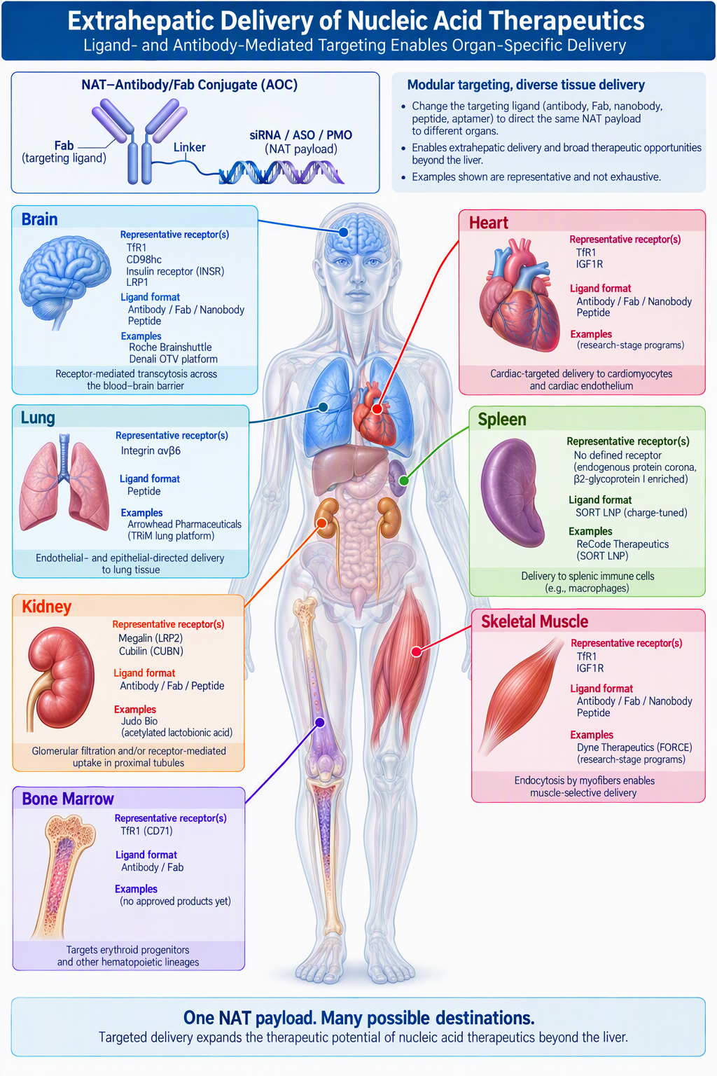
Receptor-Targeted Delivery: Recreating the GalNAc Paradigm
Every extrahepatic program now running is, in effect, a search for another ASGPR. The selection criteria are worth stating explicitly, because they separate a promising receptor from one that only looks attractive.
- High, selective expression in the target tissue. The receptor should be abundantly expressed in the target tissue to maximize tissue selectivity and minimize uptake by non-target tissues.
- Rapid, constitutive trafficking and recycling. The receptor must internalize quickly, deliver the nucleic acid therapeutic, and return to the surface quickly enough to make repeated passes. Each TfR1 molecule recycles a ligand roughly every sixteen minutes in cultured human hepatoma cells, and faster still in reticulocytes, so a receptor that is not consumed in the process can make many passes per dose.2
- No essential homeostatic role. If the receptor is essential to the health of the cell, targeting it with an antibody or ligand can cause tissue damage, particularly when the binding site overlaps that of the natural ligand.3
- Accessibility from the circulation. The receptor has to be presented on the cell surface that is directly exposed to the circulating blood.
- Conservation across species. A ligand that does not recognize the non-human primate ortholog cannot be translated to the clinic without the use of humanized animal models.
Receptors under investigation
Beyond ASGPR, the field’s work is concentrated on a short list. Transferrin receptor 1 (TfR1) is the closest thing to a general-purpose extrahepatic receptor and carries almost all the clinical-stage activity in muscle through RME. It is also an excellent shuttle across the blood-brain barrier (BBB) for delivery to the brain and the central nervous system (CNS) through receptor-mediated transcytosis (RMT). Its weakness is that it is not restricted to skeletal or cardiac muscle; it also accumulates in bone marrow, whose erythroid progenitors express more TfR1 than any other human cell type. In mice, dosing with anti-TfR1 antibodies produced acute clinical signs and a fall in circulating reticulocytes. (Reticulocytes are immature red blood cells that still carry transferrin receptors; mature red blood cells have none.) Removing Fc effector function ameliorated the adverse reaction and partly rescued the reticulocyte count, with complement accounting for the residual effect.4 Table 1 summarizes receptor targets currently under active investigation for receptor-mediated delivery, arranged approximately in order of current interest and developmental activity.
| Receptor | Major tissue(s) | Ligand/platform examples | Validation for NATs |
|---|---|---|---|
| ASGPR (ASGR1/2) | Hepatocytes | GalNAc (Alnylam, Ionis, Arrowhead, Dicerna, Novartis, Wave Life Sci. and others) | ★★★★★ |
| TfR1 (CD71) | Muscle, heart, CNS, bone marrow, proliferating cells | AOCs, FORCE (Dyne Ther.), OTV (Denali Ther.), peptides (Bicycle Ther.), aptamers (Creyon Bio) | ★★★★☆ |
| Integrin αvβ6 | Pulmonary and other epithelial tissues; carcinomas. Binds the latency-associated peptide of TGF-β (LAP-TGF-β) | Peptide-based conjugates (Arrowhead Pharm. TRiM lung platform, WO2018085415A1) | ★★★★☆ |
| No defined receptor — endogenous protein corona (β2-glycoprotein I enriched) | Spleen | SORT LNPs (ReCode Ther.); charge-tuned RNA lipoplexes | ★★☆☆☆ |
| Megalin / cubilin (LRP2 / CUBN) | Renal proximal tubule | Chemically modified carbohydrate (acetylated lactobionic acid; Judo Bio) | ★★☆☆☆ |
| GLP-1R | Pancreatic β-cells | GLP-1 receptor agonist peptide conjugates (AstraZeneca, Ionis) | ★★☆☆☆ |
| CD98hc (SLC3A2) | CNS, lung, spleen, muscle, proliferative tissues | Antibodies (Denali OTV; Roche BrainShuttle) | ★★☆☆☆ |
| IGF1R | CNS, skeletal muscle | Single-domain antibody shuttles (ABL Bio Grabody-B) | ★★☆☆☆ |
| Insulin receptor (INSR) | CNS | Antibodies | ★★☆☆☆ |
| LRP1 | CNS, neurons | Angiopep-2, melanotransferrin-derived ligands (Angiochem) | ★★☆☆☆ |
| Carbonic anhydrase IV | CNS | Brinzolamide (a carbonic anhydrase inhibitor) | ★☆☆☆☆ |
| Mannose receptor (CD206) | Macrophages and dendritic cells, including those of the spleen | Mannosylated oligos and nanoparticles | ★★☆☆☆ |
| Folate receptor α | Selected tumors | Folate conjugates | ★★☆☆☆ |
| Integrin αvβ3 | Activated and angiogenic endothelium, osteoclasts; tumor cells in melanoma and glioblastoma | Mimics of vitronectin and RGD ligands (Bicycle Ther., Philogen) | ★☆☆☆☆ |
| Integrin αvβ5 | Activated and angiogenic endothelium; epithelial tissues | Mimics of vitronectin and RGD ligands | ★☆☆☆☆ |
| EGFR | Many tumors | Peptides, aptamers, proteins | ★☆☆☆☆ |
| PSMA | Prostate cancer | PSMA ligands | ★☆☆☆☆ |
Integrin αvβ6 is the only receptor in Table 1, other than ASGPR and TfR1, for which a defined ligand-oligonucleotide conjugate has reached clinical trials. Arrowhead’s inhaled siRNA conjugates use a peptide-based αvβ6 ligand to reach pulmonary epithelium (WO2018085415A1),5 and those programs are described in the epithelial organ section below.
Megalin and cubilin offer a kidney route, and one that exploits the organ’s own scavenging machinery rather than working against it. A chemically modified carbohydrate ligand gave megalin- and cubilin-dependent uptake into proximal convoluted tubules after systemic dosing, demonstrated with antisense peptide nucleic acid conjugates that slowed fibrosis progression in a mouse model.6 Because these two receptors exist to retrieve filtered protein from the tubular lumen, they are among the few extrahepatic receptors whose physiological job is bulk endocytosis — the property that made ASGPR work.
CD98hc is the established second route across the blood-brain barrier, identified in a proteomic survey of brain endothelial cells.7 Unlike TfR1, it is internalized by a clathrin-independent route, so a conjugate that stalls on the TfR1 pathway is not necessarily stalled on this one. Carbonic anhydrase IV is the most recent entry, and was selected on brain-enrichment grounds rather than abundance alone; a small-molecule shuttle derived from an approved binder couples to proteins and oligonucleotides in a single step, and conjugates crossed the barrier in mice and neonatal macaques with parenchymal levels sustained for at least seven days.8 The in vivo work so far used antibody cargo; the oligonucleotide conjugates were taken only as far as cell-based internalization, which is why the receptor sits low in Table 1 despite the primate data.
Design guidelines for engaging these receptors
Table 2 summarizes the modalities used to engage these receptors. They form a spectrum by size, and size tracks with tissue penetration, immunogenicity risk, half-life, manufacturing cost, and whether a homogeneous conjugate can be made at all.
| Modality | Representative example | Size | NAT demonstrated | Principal trade-off |
|---|---|---|---|---|
| Lipid nanoparticles (no ligand) | SORT LNP; RNA lipoplex | ~80 nm particle | Yes (siRNA, mRNA) | Tropism set by surface charge and protein corona; cargo is encapsulated, not conjugated |
| Full IgG | anti-TfR1 AOCs (Avidity) | ~150 kDa | Yes (siRNA, PMO) | Established chemistry; long half-life; DAR heterogeneity |
| Fab | FORCE platform (Dyne Ther.) | ~50 kDa | Yes (PMO, ASO) | Smaller, no Fc effector function; shorter half-life |
| Nanobody / VHH | TfR1 VHH (Vect-Horus; Sanofi) | ~15 kDa | Yes (siRNA) | Small and stable; usually Fc-fused9 |
| Transport vehicle | OTV (Denali Ther.) | ~100 kDa | Yes (ASO) | Binding engineered into the Fc rather than the CDRs10 |
| Affibody | anti-TfR1 affibodies | ~6-7 kDa | Not yet | Small; selected to avoid the transferrin binding site; no conjugate data11 |
| Bicyclic peptide | BCY17901 (Bicycle/Ionis) | ~2 kDa | Yes (ASO, siRNA) | Fully synthetic and scalable; efficacy confirmed in non-human primates12 |
| Linear peptide | TfR1 ligand T7 peptide; Arrowhead αvβ6 ligand | ~1 kDa | Yes (siRNA) | Simplest chemistry; lower affinity, proteolytic liability13 |
| Aptamer | Anti-TfR1 aptamers (Creyon Bio) | ~10-15 kDa | Yes (ASO, siRNA) | Targeting ligand is itself an oligonucleotide; stability under optimization |
| Small molecule | CA-IV binder | <1 kDa | Cell-based only | No immunogenicity or DAR heterogeneity; ligand discovery is hard |
| Sugar / glycan | GalNAc; renal-tubule carbohydrate | <2 kDa | Yes (ASO, siRNA, PNA) | The GalNAc precedent; few receptors have a known glycan ligand |
Three design points recur across the table. First, affinity should be tuned rather than maximized. Antibodies that bind TfR1 too tightly remain associated with the brain endothelium, while lower-affinity variants are released into the parenchyma and distribute broadly;14 high-affinity engagement diverts the receptor into lysosomes instead of returning it to the surface.15 A recent single-domain antibody series makes the same point in kinetic terms: the variants that transcytosed best had intermediate affinity and fast dissociation rates rather than the tightest binding.9 Second, valency matters. Roche fused a single-chain Fab against TfR1 to the C-terminus of one or both heavy chains of an anti-amyloid-β IgG; the bivalent construct was sorted to the lysosome, whereas the monovalent version transcytosed and raised β-amyloid target engagement fifty-five-fold over the unmodified parent antibody.16 Third, the better ligands avoid the receptor’s natural ligand binding site. Hjelm et al. selected affibodies whose epitope does not overlap the transferrin site, on the reasoning that the high circulating concentration of transferrin would otherwise occupy the receptors needed for transcytosis.11
Tissue by tissue
Tissue specificity should be evaluated at four levels. The first is organ exposure, set by systemic pharmacokinetics: circulation time, clearance, and sequestration by other organs. The second is vascular access, which varies more than the rest. An antibody reaches about half its plasma concentration in the interstitium of muscle and skin — the fluid space around cells, outside the vessels — and nearly all of it in liver and spleen, where a discontinuous, or sinusoidal, endothelium leaves no real barrier between blood and tissue. In the brain it barely leaves the vessel at all.17 Where the endothelium seals the vessel, the way across is a receptor on its luminal surface, so binding precedes extravasation rather than following it. The kidney is a distinct case, because many oligonucleotides are small enough to be filtered at the glomerulus and so reach proximal tubule cells from the urinary lumen rather than across an endothelium, a route available in no other organ.
The third is cellular selectivity, within an organ as much as between organs, since almost every receptor described as tissue-enriched is expressed elsewhere too — ASGPR’s confinement to hepatocytes is the exception that made GalNAc work. The fourth is productive uptake: internalization, by a receptor or otherwise, then escape from the endosome.
Skeletal and cardiac muscle
TfR1 is expressed on myofibers and cardiomyocytes, but far more abundantly in bone marrow, where erythroid progenitors carry more of it than any other human cell type. That becomes a liability when the targeting antibody retains an intact Fc region, which engages Fcγ receptors on macrophages and opsonizes the TfR1-bearing cell for clearance; it is the source of the reticulocyte loss described above. Dyne’s FORCE platform avoids the problem by using an anti-TfR1 Fab, which has no Fc, to carry its payload (ASO, PMO, or siRNA) through a cleavable valine-citrulline linker. The linker is stable in serum but is cleaved by cathepsins in the early endosome, releasing the cargo before the conjugate reaches the lysosome.18 Avidity’s approach has instead been to engineer the Fc region of an intact anti-TfR1 antibody to be effector-silent; its lead myotonic dystrophy candidate, delpacibart etedesiran, improved the molecular pathology of the disease in the phase 1/2 MARINA study.19 Antibodies can also be dispensed with altogether. Ionis and Bicycle Therapeutics used a 2 kDa bicyclic peptide, BCY17901, that improved both ASO and siRNA potency in skeletal and cardiac muscle of human TfR1 knock-in mice, with efficacy confirmed in both tissues in non-human primates, and that avoids the conjugation heterogeneity of an antibody.12
Finally, TfR1 is classified as having low tissue specificity according to the Human Protein Atlas;20 the payload, however, adds a second layer of selectivity. The clinical candidates knock down DMPK or DUX4, or redirect dystrophin pre-mRNA splicing, and none of those has any consequence in an erythroid precursor: lowering DMPK there is pharmacologically inert, DUX4 is not expressed, and skipping exon 44 does nothing in a cell that makes no dystrophin. Therapeutic index therefore rests on three separable layers — where the conjugate distributes, where it is productively internalized, and whether the transcript is both present and consequential.
The brain and central nervous system: crossing to the other side
The brain is where vascular access dominates, since the blood-brain barrier permits almost no passive extravasation. TfR1 is again the receptor of choice. TfR1-binding ligands — antibodies, Fabs, and nanobodies — are conjugated to a NAT cargo and bind the receptor on the luminal surface of the brain endothelium. The complex is internalized and must then be sorted into a transcytotic route and released from the abluminal surface into the brain interstitium, from which the cargo diffuses to neurons and glial cells. Endothelial uptake alone is not sufficient: a substantial fraction of internalized material is retained within the endothelial cell or delivered to lysosomes, which is why the affinity, valency, and epitope rules set out above apply with particular force here. Once in the interstitial fluid, the NAT must still be taken up by the target cell and trafficked productively to reach its RNA.
Single-domain antibodies offer a smaller format, one that diffuses farther through the brain interstitium than a full IgG, although Fc fusion gives part of that advantage back.21 Camelid VHHs against TfR1 bind a novel epitope at the receptor’s dimer interface and, unlike most anti-TfR1 binders, cross-react with rodent, rhesus and human receptor, which removes the dependence on humanized animal models; in practice they are fused to an IgG1 Fc.9 Conjugated one-to-one to siRNA, they gave 50% to 80% knockdown at message and protein levels in deep brain structures of human TfR1 transgenic mice and non-human primates at doses below 1 mg/kg — structures NATs have been least able to reach at viable doses.22 One payload consideration applies whatever the shuttle: fully modified siRNA guide strands are poor substrates for cellular kinases, and a synthetic 5′-phosphate is stripped within about two hours in vivo, so a stable mimic such as 5′-(E)-vinylphosphonate is used to preserve Argonaute loading and protect the 5′ end from phosphatases and 5′-to-3′ exonucleases.23
Epithelial organs: kidney, lung, and pancreas
These three organs were each reached by a different strategy: chemistry in the kidney, route of administration in the lung, and receptor targeting in the pancreas. Grouping them emphasizes that tissue access is not solved by a single delivery technology but by exploiting the dominant biological constraint of each organ.
The kidney is often assumed to be straightforward, and it is where the usual delivery narrative breaks down. Uptake is not the limiting factor: the kidney is second only to the liver in oligonucleotide accumulation,24 even a hepatocyte-directed GalNAc ligand does not prevent retention in the renal cortex, which approaches unconjugated levels once ASGPR is saturated,25 and phosphorothioate oligonucleotides are recognized by the proximal tubule scavenging machinery, where they compete with filtered low-molecular-weight proteins for megalin- and cubilin-mediated endocytosis — the mechanism proposed for the reversible low-molecular-weight proteinuria seen in patients treated with the 2′-O-methyl phosphorothioate ASO drisapersen, which resolves during treatment interruption and occurs without histological evidence of tubular damage.26 The challenge is instead a lack of cell-type selectivity and a narrow tolerability margin. Consequently, progress has largely been driven by chemistry rather than targeting ligands. Incorporating serinol nucleic acid residues into a gapmer suppressed SGLT2 in mouse kidney more potently and durably than an unmodified DNA gapmer, with durability comparable to a 2′-O-methoxyethyl gapmer; the MOE gapmer remained the more potent of the two but caused markedly greater hepatic injury and proximal tubular injury.27 Similarly, the anti-miR-17 oligonucleotide RGLS4326 distributes preferentially to the kidney, with a kidney-to-liver exposure ratio near eight in mice,28 and raised urinary polycystin markers in a phase 1b trial in autosomal dominant polycystic kidney disease (ADPKD); development was nonetheless halted after chronic nonclinical toxicology revealed dose-limiting central nervous system toxicity, which was never seen in patients. The liability was traced to the 3′-terminal guanine, which binds the AMPA receptor ligand-binding domain in competition with glutamate; replacing it with adenine gave RGLS8429, now farabursen, which lacks the off-target interaction and has continued in clinical development.29 Receptor-based targeting through megalin and cubilin is the counterpart to these chemistry-driven approaches but remains in early stages of development. The field also bypasses renal delivery altogether when disease biology permits. Lumasiran, for example, treats primary hyperoxaluria type 1, a disorder characterized by kidney stones and progressive renal failure, by silencing glycolate oxidase in the liver.30 It is an approved therapy for a kidney disease that never acts in the kidney.
The receptors that matter depend first on whether the therapeutic can be filtered at the glomerulus. Filterable molecules, including unconjugated ASOs and siRNAs (\~7–16 kDa), encounter the proximal tubule from the urinary side, where megalin and cubilin dominate apical reabsorption.31 Ligands explored along this route include low-molecular-weight chitosan, lysozyme, and folate, all of which are filtered and then reabsorbed by the tubule; folate engages the folate receptor on the apical tubular surface.32 A sub-10 nm folate-gold nanoparticle achieved approximately 3.6% of the injected dose in a fibrotic kidney against 0.7% in the contralateral kidney.33 Because only the unbound fraction is filtered, phosphorothioate substitution, by increasing plasma protein binding, simultaneously limits glomerular filtration and prolongs systemic exposure.24
Non-filterable molecules, including antibody conjugates, Fab conjugates, and oligonucleotides engineered for high albumin binding, must approach renal targets from the vascular side. Here the glomerulus is unusually permissive: its endothelial fenestrae lack diaphragms, an architecture shared among the vascular beds surveyed only with the hepatic sinusoid, so no transcytotic step has to be engineered.34 Receptors on this side therefore function primarily in retention and cellular uptake rather than barrier crossing. Examples include VCAM-1, which has been used to direct siRNA-loaded liposomes to inflamed renal microvasculature in TNF-α-challenged mice and achieve VE-cadherin knockdown,35 the neonatal Fc receptor in podocytes,36 and α8 integrin, which is selectively expressed by mesangial cells.37 Additional targets, including CD44, kidney injury molecule-1, PDGFR-β on pericytes, and folate receptor β on profibrotic macrophages, emerge predominantly in injured tissue. A targeting ligand is not always required: an unconjugated phosphorothioate gapmer directed against APOL1 reduced target mRNA in podocytes, endothelial cells, and mesangial cells and protected against interferon-γ-induced proteinuria in APOL1-transgenic mice.38
In the lung, the key delivery decision is often made before molecular targeting is considered. Inhalation deposits the therapeutic directly onto the airway epithelium, bypassing systemic biodistribution barriers. Arrowhead’s inhaled siRNA programs, built on a platform incorporating a peptide ligand for integrin αvβ6,5 have advanced three candidates into clinical testing: one targeting the receptor for advanced glycation end products, which completed phase 1/2a evaluation and is now in a phase 2a study in allergen-induced asthma; one targeting matrix metalloproteinase 7 in idiopathic pulmonary fibrosis; and one targeting MUC5AC, which was subsequently discontinued (NCT05276570, NCT07241546, NCT05537025, NCT05292950; ClinicalTrials.gov, accessed October 5, 2026). Local administration removes much of the biodistribution challenge but not the intracellular trafficking challenge, and it restricts the strategy to diseases accessible from the airway lumen.
The pancreatic β-cell provides the clearest example of a receptor unlocking an otherwise inaccessible tissue. β-cells are notably resistant to uptake of unconjugated antisense oligonucleotides, yet conjugation to a glucagon-like peptide-1 receptor (GLP-1R) agonist produces robust gene silencing in pancreatic islets at doses that leave liver and other tissues largely unaffected.39 Structure-activity studies showed that linker design contributes as much to activity as receptor targeting itself,40 and imaging studies provided a mechanistic explanation: the oligonucleotide dissociates from the peptide early during endocytic trafficking, whereas a more stable linker prolongs co-trafficking and improves productive delivery.41 These studies remain preclinical, which is why the receptor sits at in vivo proof of concept in Table 1.
The spleen: selectivity without a ligand
Like the liver, the spleen is served by discontinuous sinusoidal endothelium that leaves no real barrier between blood and tissue, and an antibody reaches nearly its full plasma concentration in the splenic interstitium.17 The difficulty is that the same architecture serves the liver, which is larger, more heavily perfused, and equipped with a receptor system that captures lipid particles avidly. Splenic delivery is therefore a problem of selectivity against the liver rather than of access, and the approaches that work do not use a targeting ligand at all. Instead, a very different strategy has shown remarkable success – the use of lipid nanoparticles (LNPs). The clearest demonstration is the selective organ targeting (SORT) platform by ReCode Therapeutics. It adds a fifth lipid to a conventional four-component lipid nanoparticle and tunes spleen tropism through its charge and molar fraction. Incorporating 10% to 40% of the anionic lipid 1,2-dioleoyl-sn-glycero-3-phosphate, the LNP redirected reporter expression entirely to the spleen, with no signal detected in any other organ; the same design with a permanently cationic lipid delivers to lung, and with an ionizable cationic lipid to liver.42 The mechanism is as follows: Poly(ethylene glycol) lipids desorb in circulation, the exposed targeting lipid recruits a distinct set of plasma proteins, and those proteins engage receptors enriched in the destination tissue.43 For the spleen the enriched protein is β2-glycoprotein I, although no cognate splenic receptor has been identified and its functional role is described by the authors as suggested rather than established.
Silencing data followed only recently, and they are more modest than the biodistribution implies. Spleen-directed SORT particles carrying siRNA reduced the endogenous transcript Pecam1 by approximately 57% in mouse spleen after a single intravenous dose, and by approximately 67% at both message and protein after two doses of 1.5 mg/kg. Resolved by cell type, splenic endothelial and immune populations showed roughly 18% to 20% silencing, and the splenic formulation was the least potent of the three organ-directed designs in that study.44 The spleen therefore sits apart from every other tissue in this review. Elsewhere a ligand supplies the address. Here tropism follows from the surface charge of a particle and the plasma proteins that charge recruits, and the cargo must be encapsulated rather than conjugated. No spleen-directed lipid nanoparticle has entered clinical testing for gene silencing; the clinical work on this route so far is vaccination, not knockdown.
What the tissues have in common
No tissue was solved by a targeting ligand alone, and only one was solved by chemistry alone. Each first required a solution to tissue access: a borrowed receptor in muscle, receptor-mediated transcytosis at the blood-brain barrier, local administration in the lung, receptor targeting in pancreatic β-cells, and in the spleen a particle surface that recruits the right plasma proteins. Chemistry then enabled the oligonucleotide to survive, distribute, and function within the compartment that strategy made accessible. The kidney and the spleen are the exceptions, and they are exceptions of the same kind. Both are reached without help, the kidney through glomerular filtration and the spleen through sinusoidal endothelium, and in both the limiting problem was never access but selectivity: among cell types in the kidney, and against the liver in the spleen. The pattern is ultimately the same as in the liver, except that the liver supplies tissue access almost automatically through its physiology.
Tissue exposure is not the endpoint, and it is one of the most frequently misinterpreted measurements in the field. Increasing phosphorothioate content in a cholesterol-conjugated siRNA substantially increased cellular uptake without improving silencing, because much of the additional material accumulated in endosomes and lysosomes rather than the cytosol, where the RNA-induced silencing complex assembles.45 Similarly, knockdown of a ubiquitously expressed transcript in muscle by antibody-siRNA conjugates plateaued above 1 mg/kg even as tissue concentrations continued to rise,46 while improvements in newer GalNAc designs were driven primarily by enhanced metabolic stability rather than more efficient receptor engagement.47 These observations point to the same bottleneck. An oligonucleotide may reach the correct tissue and even enter the correct cell, yet remain pharmacologically inactive if it cannot escape the endosomal pathway. Biodistribution determines where a therapeutic goes; endosomal escape determines whether it works. The two challenges are largely orthogonal, and it is endosomal escape that now defines the next frontier of extrahepatic oligonucleotide delivery.
References
- Egli M, Manoharan M. Chemistry, structure and function of approved oligonucleotide therapeutics. Nucleic Acids Res. 2023;51(6):2529-2573. doi:10.1093/nar/gkad067
- Ciechanover A, Schwartz AL, Dautry-Varsat A, Lodish HF. Kinetics of internalization and recycling of transferrin and the transferrin receptor in a human hepatoma cell line. Effect of lysosomotropic agents. J Biol Chem. 1983;258(16):9681-9689.
- Haqqani AS, Belanger K, Stanimirovic DB. Receptor-mediated transcytosis for brain delivery of therapeutics: receptor classes and criteria. Front Drug Deliv. 2024;4:1360302. doi:10.3389/fddev.2024.1360302
- Couch JA, Yu YJ, Zhang Y, et al. Addressing safety liabilities of TfR bispecific antibodies that cross the blood-brain barrier. Sci Transl Med. 2013;5(183):183ra57. doi:10.1126/scitranslmed.3005338
- Anand P, Zhang Y, Patil S, Kaur K. Metabolic stability and targeted delivery of oligonucleotides: advancing RNA therapeutics beyond the liver. J Med Chem. 2025;68(7):6870-6896. doi:10.1021/acs.jmedchem.4c02528
- Kumar V, Wahane A, Tham MS, et al. Efficient and selective kidney targeting by chemically modified carbohydrate conjugates. Mol Ther. 2024;32(12):4383-4400. doi:10.1016/j.ymthe.2024.10.020
- Zuchero YJY, Chen X, Bien-Ly N, et al. Discovery of novel blood-brain barrier targets to enhance brain uptake of therapeutic antibodies. Neuron. 2016;89(1):70-82. doi:10.1016/j.neuron.2015.11.024
- Ding X, Chen X, Boehm P, et al. CA-IV-directed small-molecule shuttle enables targeted brain delivery of biologics. Nat Chem Biol. Published online August 26, 2026. doi:10.1038/s41589-026-02294-y
- David M, Cohen R, Beuzelin D, et al. Novel single-domain antibodies targeting a unique transferrin receptor 1 epitope for cross-species delivery of drugs in the central nervous system. J Control Release. 2025;388(Pt 2):114344. doi:10.1016/j.jconrel.2025.114344
- Barker SJ, Thayer MB, Kim C, et al. Targeting the transferrin receptor to transport antisense oligonucleotides across the mammalian blood-brain barrier. Sci Transl Med. 2024;16(760):eadi2245. doi:10.1126/scitranslmed.adi2245
- Hjelm LC, Lindberg H, Ståhl S, Löfblom J. Affibody molecules intended for receptor-mediated transcytosis via the transferrin receptor. Pharmaceuticals (Basel). 2023;16(7):956. doi:10.3390/ph16070956
- Østergaard ME, Carrer M, Anderson BA, et al. Conjugation to a transferrin receptor 1-binding Bicycle peptide enhances ASO and siRNA potency in skeletal and cardiac muscles. Nucleic Acids Res. 2025;53(7):gkaf270. doi:10.1093/nar/gkaf270
- Fu T, Fan F, Lin Y, et al. TfR1-binding peptide conjugation facilitates robust and specific siRNA delivery to the central nervous system. Bioconjug Chem. 2025;36(7):1377-1383. doi:10.1021/acs.bioconjchem.5c00225
- Yu YJ, Zhang Y, Kenrick M, et al. Boosting brain uptake of a therapeutic antibody by reducing its affinity for a transcytosis target. Sci Transl Med. 2011;3(84):84ra44. doi:10.1126/scitranslmed.3002230
- Bien-Ly N, Yu YJ, Bumbaca D, et al. Transferrin receptor (TfR) trafficking determines brain uptake of TfR antibody affinity variants. J Exp Med. 2014;211(2):233-244. doi:10.1084/jem.20131660
- Niewoehner J, Bohrmann B, Collin L, et al. Increased brain penetration and potency of a therapeutic antibody using a monovalent molecular shuttle. Neuron. 2014;81(1):49-60. doi:10.1016/j.neuron.2013.10.061
- Eigenmann MJ, Karlsen TV, Krippendorff BF, et al. Interstitial IgG antibody pharmacokinetics assessed by combined in vivo- and physiologically-based pharmacokinetic modelling approaches. J Physiol. 2017;595(24):7311-7330. doi:10.1113/JP274819
- Weeden T, Picariello T, Quinn B, et al. FORCE platform overcomes barriers of oligonucleotide delivery to muscle and corrects myotonic dystrophy features in preclinical models. Commun Med (Lond). 2025;5(1):22. doi:10.1038/s43856-025-00733-w
- Kwan TT, Meng Q, Delos Santos N, et al. Delpacibart etedesiran improves the molecular pathology of myotonic dystrophy type 1 in the phase 1/2 MARINA study. Mol Ther. 2026;34(7):3872-3884. doi:10.1016/j.ymthe.2026.03.013
- TFRC. The Human Protein Atlas. Accessed October 6, 2026. https://www.proteinatlas.org/ENSG00000072274-TFRC
- Thorne RG, Nicholson C. In vivo diffusion analysis with quantum dots and dextrans predicts the width of brain extracellular space. Proc Natl Acad Sci U S A. 2006;103(14):5567-5572. doi:10.1073/pnas.0509425103
- Jacquot G, David M, Pecqueux B, et al. Harnessing TfR1 for cross-species systemic delivery of siRNAs to deep brain regions using single-domain antibodies. bioRxiv. Preprint posted online May 22, 2026. doi:10.64898/2026.05.20.726486
- Haraszti RA, Roux L, Coles AH, et al. 5′-Vinylphosphonate improves tissue accumulation and efficacy of conjugated siRNAs in vivo. Nucleic Acids Res. 2017;45(13):7581-7592. doi:10.1093/nar/gkx507
- Geary RS, Norris D, Yu R, Bennett CF. Pharmacokinetics, biodistribution and cell uptake of antisense oligonucleotides. Adv Drug Deliv Rev. 2015;87:46-51. doi:10.1016/j.addr.2015.01.008
- Sewing S, Gubler M, Gérard R, et al. GalNAc conjugation attenuates the cytotoxicity of antisense oligonucleotide drugs in renal tubular cells. Mol Ther Nucleic Acids. 2019;14:67-79. doi:10.1016/j.omtn.2018.11.005
- Janssen MJ, Nieskens TTG, Steevels TAM, et al. Therapy with 2′-O-Me phosphorothioate antisense oligonucleotides causes reversible proteinuria by inhibiting renal protein reabsorption. Mol Ther Nucleic Acids. 2019;18:298-307. doi:10.1016/j.omtn.2019.08.025
- Tsuboi T, Hattori K, Ishimoto T, et al. In vivo efficacy and safety of systemically administered serinol nucleic acid-modified antisense oligonucleotides in mouse kidney. Mol Ther Nucleic Acids. 2025;36(1):102387. doi:10.1016/j.omtn.2024.102387
- Lee EC, Valencia T, Allerson C, et al. Discovery and preclinical evaluation of anti-miR-17 oligonucleotide RGLS4326 for the treatment of polycystic kidney disease. Nat Commun. 2019;10(1):4148. doi:10.1038/s41467-019-11918-y
- Valencia T, Yen LY, Berman C, et al. The nucleobase guanine at the 3′-terminus of oligonucleotide RGLS4326 drives off-target AMPAR inhibition and CNS toxicity. Nat Commun. 2025;16(1):10762. doi:10.1038/s41467-025-65799-5
- Garrelfs SF, Frishberg Y, Hulton SA, et al. Lumasiran, an RNAi therapeutic for primary hyperoxaluria type 1. N Engl J Med. 2021;384(13):1216-1226. doi:10.1056/NEJMoa2021712
- Christensen EI, Birn H. Megalin and cubilin: multifunctional endocytic receptors. Nat Rev Mol Cell Biol. 2002;3(4):256-266. doi:10.1038/nrm778
- Zhou P, Sun X, Zhang Z. Kidney-targeted drug delivery systems. Acta Pharm Sin B. 2014;4(1):37-42. doi:10.1016/j.apsb.2013.12.005
- Chan CKW, Szeto CC, Lee LKC, et al. A sub-10-nm, folic acid-conjugated gold nanoparticle as self-therapeutic treatment of tubulointerstitial fibrosis. Proc Natl Acad Sci U S A. 2023;120(42):e2305662120. doi:10.1073/pnas.2305662120
- Stan RV, Tse D, Deharvengt SJ, et al. The diaphragms of fenestrated endothelia: gatekeepers of vascular permeability and blood composition. Dev Cell. 2012;23(6):1203-1218. doi:10.1016/j.devcel.2012.11.003
- Kowalski PS, Zwiers PJ, Morselt HWM, et al. Anti-VCAM-1 SAINT-O-Somes enable endothelial-specific delivery of siRNA and downregulation of inflammatory genes in activated endothelium in vivo. J Control Release. 2014;176:64-75. doi:10.1016/j.jconrel.2013.12.029
- Dylewski J, Dobrinskikh E, Lewis L, et al. Differential trafficking of albumin and IgG facilitated by the neonatal Fc receptor in podocytes in vitro and in vivo. PLoS One. 2019;14(2):e0209732. doi:10.1371/journal.pone.0209732
- Hartner A, Schöcklmann H, Pröls F, Müller U, Sterzel RB. Alpha8 integrin in glomerular mesangial cells and in experimental glomerulonephritis. Kidney Int. 1999;56(4):1468-1480. doi:10.1046/j.1523-1755.1999.00662.x
- Aghajan M, Booten SL, Althage M, et al. Antisense oligonucleotide treatment ameliorates IFN-γ-induced proteinuria in APOL1-transgenic mice. JCI Insight. 2019;4(12):e126124. doi:10.1172/jci.insight.126124
- Ämmälä C, Drury WJ 3rd, Knerr L, et al. Targeted delivery of antisense oligonucleotides to pancreatic β-cells. Sci Adv. 2018;4(10):eaat3386. doi:10.1126/sciadv.aat3386
- Knerr L, Prakash TP, Lee R, et al. Glucagon like peptide 1 receptor agonists for targeted delivery of antisense oligonucleotides to pancreatic beta cell. J Am Chem Soc. 2021;143(9):3416-3429. doi:10.1021/jacs.0c12043
- Kay E, Stulz R, Becquart C, et al. NanoSIMS imaging reveals the impact of ligand-ASO conjugate stability on ASO subcellular distribution. Pharmaceutics. 2022;14(2):463. doi:10.3390/pharmaceutics14020463
- Cheng Q, Wei T, Farbiak L, Johnson LT, Dilliard SA, Siegwart DJ. Selective organ targeting (SORT) nanoparticles for tissue-specific mRNA delivery and CRISPR-Cas gene editing. Nat Nanotechnol. 2020;15(4):313-320. doi:10.1038/s41565-020-0669-6
- Dilliard SA, Cheng Q, Siegwart DJ. On the mechanism of tissue-specific mRNA delivery by selective organ targeting nanoparticles. Proc Natl Acad Sci U S A. 2021;118(52):e2109256118. doi:10.1073/pnas.2109256118
- Vaidya A, Moore S, Chatterjee S, et al. Expanding RNAi to kidneys, lungs, and spleen via selective organ targeting (SORT) siRNA lipid nanoparticles. Adv Mater. 2024;36(35):e2313791. doi:10.1002/adma.202313791
- Ly S, Echeverria D, Sousa J, Khvorova A. Single-stranded phosphorothioated regions enhance cellular uptake of cholesterol-conjugated siRNA but not silencing efficacy. Mol Ther Nucleic Acids. 2020;21:991-1005. doi:10.1016/j.omtn.2020.07.029
- Cochran M, Arias D, Burke R, et al. Structure-activity relationship of antibody-oligonucleotide conjugates: evaluating bioconjugation strategies for antibody-siRNA conjugates for drug development. J Med Chem. 2024;67(17):14852-14867. doi:10.1021/acs.jmedchem.4c00802
- Foster DJ, Brown CR, Shaikh S, et al. Advanced siRNA designs further improve in vivo performance of GalNAc-siRNA conjugates. Mol Ther. 2018;26(3):708-717. doi:10.1016/j.ymthe.2017.12.021